Low back pain most often affects people after the age of 35. In the vast majority of cases, the disease is associated with the deformity of the vertebrae and its consequences. A timely visit to a doctor will speed up recovery, because the symptoms and treatment of osteochondrosis of the lumbar spine are interrelated concepts.
The more the disease progresses, the more serious its consequences and the more difficult the process of recovering health.
Signs and symptoms of osteochondrosis of the lumbar spine
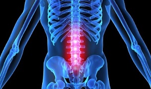
The lumbar spine is located between the sacrum and the thoracic region and consists of five vertebrae connected by intervertebral discs.
The development of osteochondrosis means the wear of the intervertebral discs, which play a cushioning role during loads on the spine. The base of the discs is a gelatinous mass, protected by a dense fibrous annulus and cartilaginous tissue, and the interior space is filled with a liquid nucleus pulposus.
As the loads on the vertebrae increase, the elasticity and flexibility of the intervertebral discs, as well as their height, are lost, and microcracks form in the annulus fibrosus, eventually leading to its rupture and damage to the nucleus pulposus.
Tissue destruction is accompanied by pinching of the nerve roots located on both sides of the vertebrae and causes severe pain.
The main signs of lumbar osteochondrosis:
- back pain;
- fatigue and depression;
- excessive muscle weakness or tension;
- loss of sensation in the extremities, buttocks or thighs;
- sharp or aching pains and spasms in the lower back, often radiating to the legs;
- violation of motor function.
Against the background of severe injuries of the vertebrae in the lumbar region, other symptoms are observed, most often dysfunctions of other organs: the urinary and reproductive systems, gastrointestinal tract.
Causes of occurrence
Like most diseases of the musculoskeletal system, osteochondrosis can develop for many reasons. Some of them have their roots in lifestyle and diet, while the other part develops against the background of the physiological characteristics of the body.
Very often, athletes require the treatment of osteochondrosis of the lumbosacral spine, the back of which is exposed not only to constant power loads, but also to periodic injuries.

The second category of people at risk, people who, by virtue of their profession, spend a lot of time in a position: teachers, hairdressers, cooks, porters, waiters, programmers, office workers and drivers.
Among other reasons for the development of pathology:
- overweight;
- metabolic disorder;
- wrong posture, stooping;
- genetic predisposition;
- lesions;
- bad habits;
- lack of useful trace elements and vitamins in the diet;
- abnormal development of the musculoskeletal system, flat feet;
- hypothermia;
- idle, static;
- frequent stress.
All these factors can affect the elasticity of the intervertebral discs, since they contribute to the alteration of blood circulation or the appearance of a deficiency of nutrients that enter the vertebral tissues.
The vertebrae are capable of carrying out their functions, subject to regular tissue renewal. In case of malnutrition of the vertebral tissues, either due to lack of blood circulation or problems with metabolism, the regeneration processes slow down or stop completely. Then there is a dystrophic and desiccation change in the cartilage and the fibrous ring of the vertebrae.
Degrees of osteochondrosis of the lumbar spine
Depending on the level of the spinal injury, there are four degrees of development of osteochondral processes, which manifest in stages, as the disease progresses.
First grade
Pathological processes in the spine begin long before their first clinical manifestation. As a result of the loss of moisture, the intervertebral discs become less elastic. The height of the discs remains normal. The patient feels discomfort in the lumbar region.
Second grade
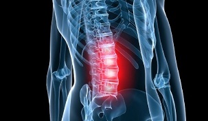
In the context of moisture deficiency, microcracks appear in the annulus fibrosus and inflammation of the tissues develops.
The hooked processes of the vertebrae gradually increase. The seals develop in the cartilage.
A patient complains of back pain that radiates to the legs or groin. Limitation of motor skills is possible. Failures occur in the work of internal organs.
Third degree
The integrity of the annulus fibrosus is compromised, the intervertebral disc protrudes and forms a hernia. The vessels and nerve endings are compressed. There are muscle spasms, dysfunction of the pelvic organs, sensory disorder of the lower extremities, prolonged attacks of sciatica.
Fourth degree
The most difficult, non-treatable stage in the course of the disease. As a result of the complete destruction of the intervertebral discs, scars are formed in their place. The vertebrae get as close as possible and gradually deform. With the development of compression of the spinal cord, paralysis of the lower extremities is possible.
If timely treatment of osteochondrosis of the lumbar spine is not provided, the destruction of the vertebrae will progress and may lead to disability.
Diagnosis
To recognize a disease and determine an accurate diagnosis, neurologists use a set of measures: anamnesis, physiological examination, and instrumental studies.
Taking anamnesis
Provides for the study of patient complaints:
- cause for concern;
- location of discomfort;
- duration and intensity of unpleasant sensations;
- the duration of the disease;
- possible causes of the disease;
- frequency of exacerbations;
- factors causing exacerbations;
- factors that improve well-being.
In addition, the doctor studies information about the patient's lifestyle, diet, work and rest, the presence of bad habits, hereditary factors and trauma.
Physiological examination

Performed to identify pathological changes and make a preliminary diagnosis.
During the exam, the doctor assesses the patient's motor skills: gait, posture, range, and range of motion. The method of palpation examines the state of the muscles: tone, size, volume, presence of spasms.
Sets the sensitivity level with a slight tingling sensation. Hitting with a hammer makes it possible to recognize the areas of radiation of pain.
Hardware Studies
To obtain complete and accurate information on the location of the pathology and the degree of tissue damage, doctors use research with various types of medical equipment.
Radiography.Examination of the lumbar spine by radiography allows to establish the anatomical parameters of the vertebrae and intervertebral discs, the tendency to narrow the holes between the bases, the presence of bone growths.
Tomography.The use of electromagnetic waves provides an image of the area under study on the screen for further study and analysis of the state of the vessels that supply the tissues of the spine, the nervous processes and the intervertebral discs.
CT.X-ray images are taken of various segments of the spine. The image is displayed on the monitor to determine the nature of changes in the vessels, the membrane of the vertebrae and the spinal cord, marginal growths.
For differential diagnosis, various types of investigation are used to exclude pathologies of other body systems.
Treatment of osteochondrosis of the lumbosacral spine
The duration and characteristics of the treatment of lumbosacral osteochondrosis depend on the results of the diagnostic measures. In the early stages of the development of the disease, conservative treatment is indicated. For more complex injuries to the spine, surgical intervention is used.
The optimal therapeutic effect is achieved by complex therapy, which includes the use of topical medications, physical therapy, massage and gymnastics to improve health.
Medications
To relieve symptoms, non-steroidal drugs are prescribed for internal and external use: tablets, injections, ointments. In addition, chondroprotectors, neuroprotectors, diuretics, vitamins, muscle relaxants are used.

Medication allows:
- eliminate pain;
- relieve inflammation;
- relax muscles;
- restore destroyed cartilage tissue;
- improve blood circulation;
- reduce swelling;
- increase physical activity;
- normalizes brain nutrition.
For acute pain, the novocaine blockade is used, which provides instant action.
Folk remedies
Traditional treatment is effective as an adjunct to drug therapy. The main methods of traditional medicine are based on the use of plant materials, animal products and chemicals.
On the basis of various components, ointments and compresses, decoctions and infusions are prepared, which are used for internal and external use, as well as for therapeutic baths.
Physiotherapy for lumbar osteochondrosis
Physical therapy procedures are an excellent way to restore motor functions in the spine after suffering from osteochondrosis.
The main physiotherapy methods are:
- electrotherapy: exposure to weak electrical currents to improve blood circulation in tissues;
- magnetotherapy: the use of the properties of the magnetic field to restore tissue at the cellular level;
- laser therapy: complex activation of biological processes in vertebral tissues and nerve endings;
- shock wave therapy: improvement of microcirculation and metabolic processes in tissues affected by exposure to an acoustic wave;
- balneotherapy- using the healing properties of mineral water.
Physiotherapy procedures not only increase the effectiveness of drug treatment several times, but also contribute to the healing and strengthening of the body as a whole.
Massage for osteochondrosis of the lumbar region
Visiting massage treatments is one of the most pleasant and effective methods of treating osteochondrosis.
With massage therapy:
- eliminate muscle spasms;
- improve blood supply to affected areas;
- improve lymphatic flow;
- restores atrophied muscles;
- remove mobility limitation.
Massage is prescribed when pain syndromes are eliminated.
Recovery gymnastics
The main task of exercise therapy for osteochondrosis is the restoration of the functionality of the spine and its correction. However, you can attend classes only after eliminating the symptoms of exacerbation.
The most effective methods of medical gymnastics are:
- loading;
- visit to the gym;
- water therapy, swimming.
A hoop can be used to play sports at home. Some doctors recommend yoga to their patients to restore flexibility in the spine.
Exercises for exacerbation of lumbar osteochondrosis
Any exercise for osteochondrosis should be done slowly and without sudden movements.
To strengthen the muscles that support the vertebrae, appropriate exercises are performed while lying on your stomach. In this case, the arms are raised with a slight stretch, but without tension. Repeat 4 times.
Surgery
Surgery is used to treat the spine in especially difficult cases, with significant neurological disorders, as well as loss of control over stool.
During surgery, the source of disease is removed and steps are taken to stabilize the spine. The postoperative period lasts several months.
Why is lumbar osteochondrosis dangerous?
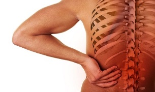
The degenerative changes that occur in lumbar osteochondrosis contribute to the development of many life-threatening diseases. In the context of an intervertebral hernia, lumps, lumbago and sciatica occur.
Further progression of the disease can lead to intervertebral disc prolapse and spinosis formation. In addition to the intense pain that accompanies the pathology, the motor abilities of a person are interrupted, until their complete loss. Paralysis of the lower extremities develops.
Death is inevitable with significant damage to the lining of the spinal cord.
Prevention
To avoid harmful changes in the spine, you must take care of a healthy lifestyle:
- practices sports: swimming, temperate;
- to adhere to a correct balanced and nutritious diet;
- eliminate bad habits;
- hold posture;
- support the spine during sleep with an orthopedic mattress.
Also, it is advisable to avoid hypothermia, lifting heavy objects. Women are advised not to wear high heels frequently.
You can keep your lower back healthy by adjusting your lifestyle and not forgetting the importance of physical activity.


